The 4-year male child was brought with complaints of passing thin stream of urine from the meatus and from the coronal region. He underwent tubularized incised plate urethroplasty 1 year ago at 3 years of age. On clinical examination there was coronal fistula with narrow meatus at glans. Prepucial skin was absent since it was used during urethroplasty in the first surgery. He had good glans groove. In view of the above findings, we planned for cystoscopy and meatotomy/urethroplasty. Oral mucosa inlay graft in failed hypospadias was considered as part of the treatment plan.
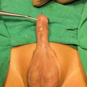
Fig 1: Clinical picture showing coronal fistula
Cystoscopy was done to delineate the length of abnormal urethra, specifically targeting “Oral mucosa inlay graft in failed hypospadias.” Cystoscopy done using 4.5Fr scope since the meatus was very narrow. Narrow urethra noted up to the corona. Rest of the urethra appeared normal. Hence decided to proceed with distal urethroplasty.
Stay suture taken over the glans and marking done to lay open the tract upto the fistula. Local anaesthesia was infiltrated at the marked incision site. Urethra laid open upto the fistula site. Urethral plate incised in the midline and the edges drawn apart. We planned to place a small oral mucosa graft to widen the urethral plate.
Oral mucosa graft harvest:
Oral mucosa graft harvested from the upper lip to the right of the frenulum. Stay suture taken over the vermilion border. Incision marked and local anaesthesia infiltrated at the marked site. 2x1cm graft harvested and defatted.
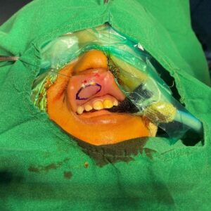
Fig 2: Oral mucosa graft marking
This oral mucosa graft was placed in the diamond shaped defect and sutured all around at the edges. In addition to this the graft was quilted in the centre into the neourethral groove. Following this the urethral plate was tubularized over a 7Fr Infant feeding tube using 6-0 PDS in a continuous subcuticular pattern. Local tissues were used as a second layer.
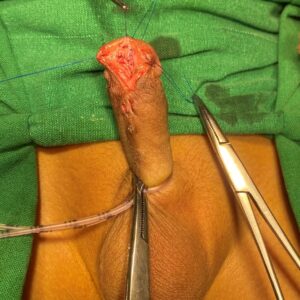
Fig 3: Oral mucosa graft placed in the defect to widen the urethral plate and sutured at the edges and quilted in the centre.
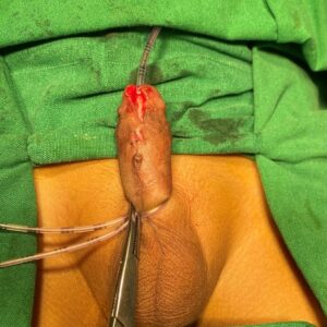
Fig 4: Completion of first layer of urethroplasty using 6-0 PDS
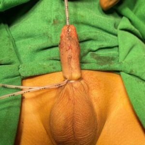
Fig 5: Urethroplasty done over a 7Fr Infant Feeding tube.
Dressing was removed on post-operative day 7. Catheter was removed on post-operative day 14. Following catheter removal, he was passing urine from the neomeatus with no complications.
At post-operative follow up at 1 month the meatus was healthy, urine stream good with no complications.
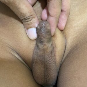
Fig 6: Post operative follow up at 1 month
If you wish to contact him, pls fill up this form- Contact form for Dr Singal
or pls call up his clinic for an appointment- Clinic details for Dr Singal.
